Table of Contents
Structural and Functional Imaging Techniques with the 4T Scanner
Overview of the BIAC 4T Scanner The 4 Tesla scanner is a passively shielded GE LX NVi system. Like the 3 T scanner, it is equipped with a high power 41-mT/m gradient system and four-channel parallel receivers at 1MHz sampling frequency that enable single-shot echo-planar and spiral imaging. It uses broadband transmission to enable multiple frequency excitation schemes, which are important for multi-nuclei imaging and spectroscopy experiments. To ensure high homogeneity critical for fMRI experiments at high field, the 4 T scanner is equipped with high-order room-temperature resistive shimming coils in addition to the super-conducting shimming coils.
Structural Imaging Techniques
The 4T scanner uses several main types of structural imaging techniques: T1 Imaging, T2-weighted coplanar and 2D Proton Density.
- T1 Imaging
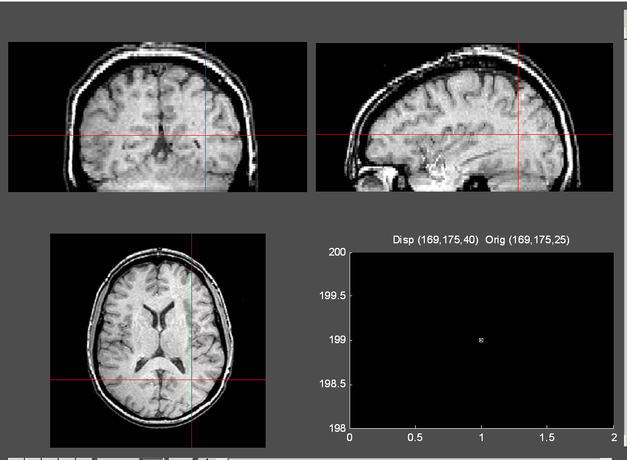
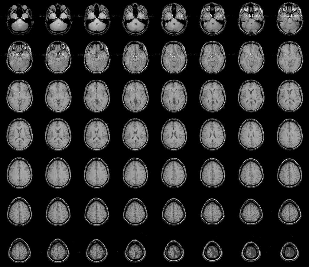
T1 Imaging provides information about the T1 values of tissue. A typical T1 contrast is generated using short TEs and moderate TRs. A common T1 imaging sequence is based on a 3D Fast Spoiled Gradient Recalled (FSPGR) acquisition. The representative imaging parameters for 3D FSPGR acquisition to achieve T1 contrast are as follow:- TE 5.4ms, TR 12.3s, Flip angle 20°
- Imaging matrix 256×160, FOV 24 cm
- Slice thickness 1.9mm, 72 slices
- Number of Excitation (NEX) 1
- T2 Imaging
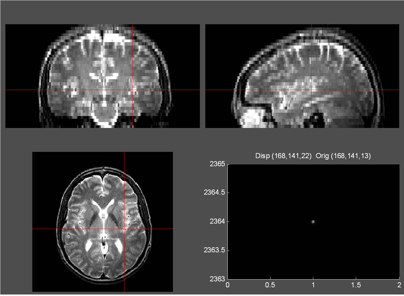
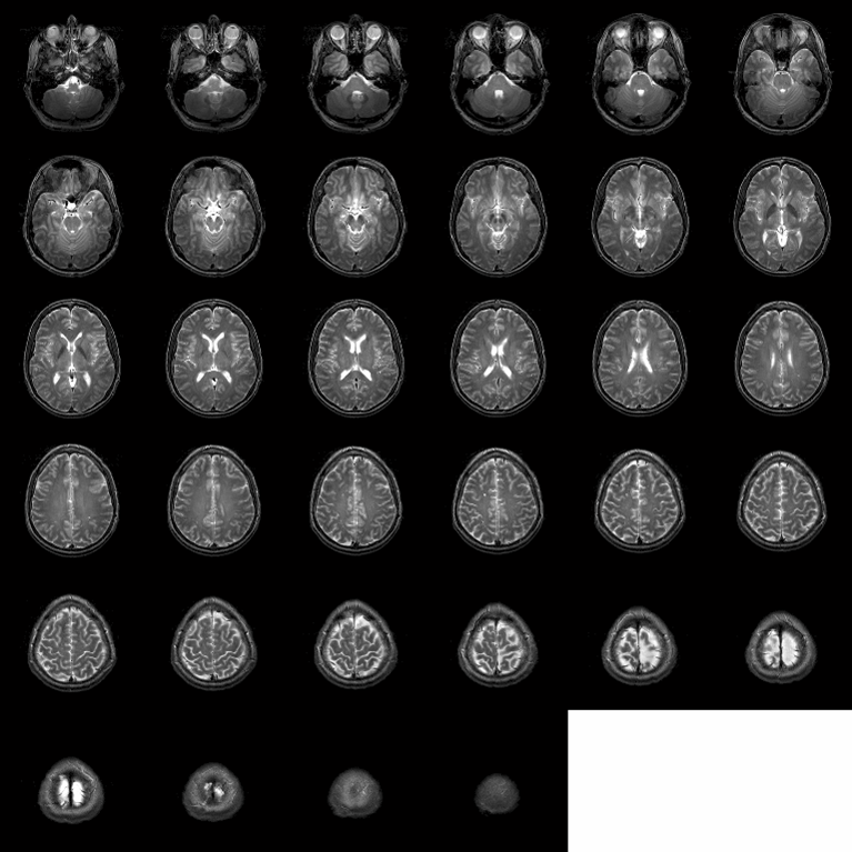
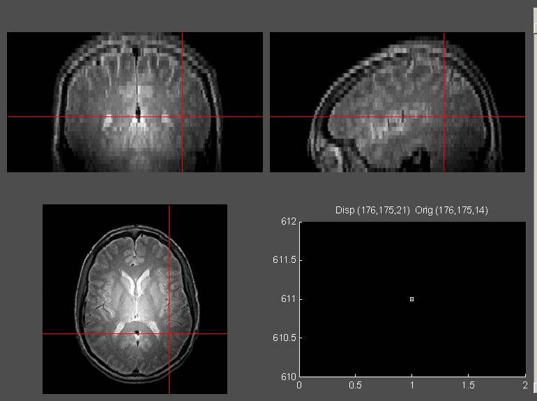
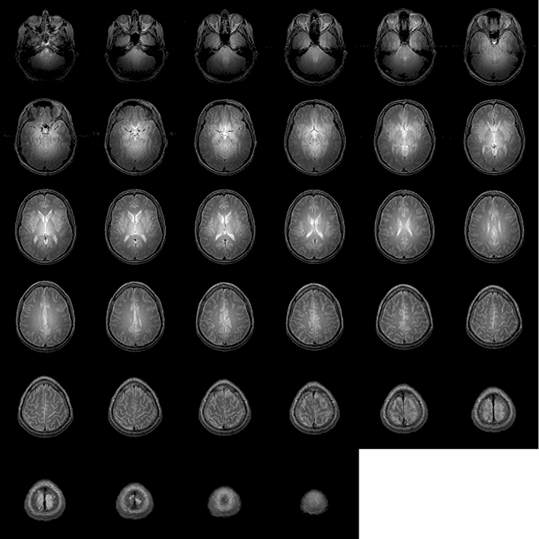
T2-weighted coplanar imaging provides information about the relative T2 values of tissue. T2 contrast is typically generated using moderate TEs and long TRs. A common T2 weighted imaging sequence is based on a 2-D Fast Spin Echo pulse sequence. The representative imaging parameters for 2D FSE acquisition to achieve T2 contrast are as follow:- 2D Fast Spin Echo (FSE)
- TE 68ms, TR 9s, echo train length 4
- Imaging matrix 256×192, FOV 24 cm
- Slice thickness 3.8mm, 34 slices
- Number of Excitation (NEX) 1
- 2-D Proton Density Imaging
Proton-Density Imaging creates MR images that are sensitive to the number of protons present within each voxel. Proton-Density Images are typically acquired with short TEs and long TRs. A 2-D Fast Spin Echo pulse sequence can also be used to obtain PD contrast with the following parameters:- 2D Fast Spin Echo (FSE)
- TE 17 ms, TR 6 s, echo train length 4
- Imaging matrix 256×256, FOV=24
- Slice thickness 3.8mm, 34 slices
- Number of Excitation (NEX) 1
Functional Sequences on the 4T
Functional sequences are designed to map out patterns of activity at different regions of the brain. BIAC built functional sequences upon the spiral waveform originated from Stanford (Dr. Gary Glover) and echo-planar waveform originated from MCW (Dr. Eric Wong currently at UCSD). Spiral imaging is a technique for fast image acquisition that uses sinusoidally changing gradients to trace a corkscrew trajectory through k-space. K-space is a notation scheme used to describe MRI data. Echo-planar imaging (EPI), is a technique that allows for the collection of an entire two-dimensional image by changing spatial gradients rapidly following a single electromagnetic pulse from a transmitter coil.
- Duke EPI
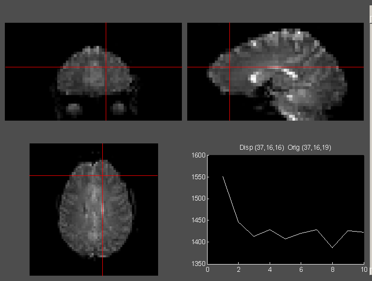
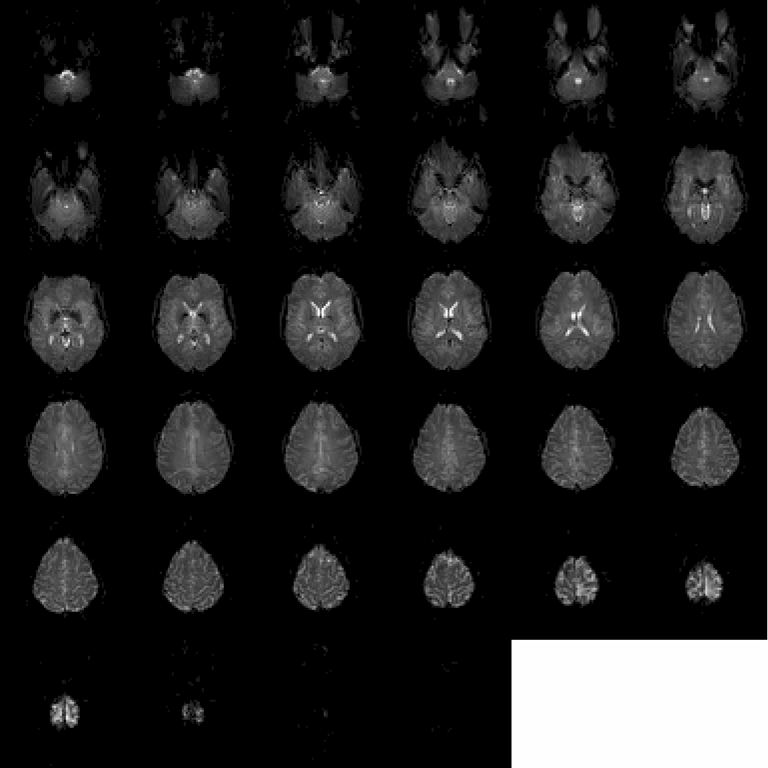
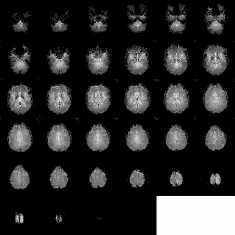
Duke EPI, an echo-planar imaging technique developed in-house at BIAC, allows for the collection of an entire two-dimensional image by changing spatial gradients rapidly following a single electromagnetic pulse from a transmitter coil. The typical imaging parameters are as follow:- TE 30 ms, TR 2s, Flip angle 60°
- Slice thickness 3.8mm
- 34 slices
- Spiral-In
Spiral imaging sequences use sinusoidal changes in the gradients to trace a corkscrew path through k-space. Spiral-In Imaging follows a corkscrew path that beings at the periphery of k-space and ends at the center. The typical imaging parameters are as follow:- TE 30 ms, TR 2s, Flip angle 60°
- Slice thickness 3.8mm
- 34 slices
Functional Sequence Summary
Each of these techniques has its advantages and its disadvantages:
- The Spiral-In technique has better signal recovery at the ventral brain region than the spiral-out technique, and it is capable of high throughput. Spiral-in imaging, however, has blurry edges and low internal contrast.
- EPI allows for both high internal contrast and clear image edges, but it has spatial distortions at the ventral and frontal brain regions, along with some signal losses.
