Table of Contents
BIAC MRI Scanners
BIAC’s major instruments include a 3.0 Tesla GE EXCITE HD whole-body 60 cm bore human scanner, a 3.0 Tesla (T) GE MR750 whole-body 60 cm bore human scanner, a 7.0 Tesla Bruker BioSpec 33 cm bore animal scanner, and mock scanner facilities.
Current Scanners
GE MR750 3T Scanner (BIAC5 & BIAC6)
The 3.0 T GE MR750 scanner uses an actively shielded magnet design to restrict fringe magnetic fields. This scanner is equipped with a 32-channel parallel receiving architecture (expandable to 128 channels), an 8-channel head coil, 32-channel head coil, and a 16-Channel Head/Neck/Spine Array. The scanner is equipped with a high-powered gradient system that has a maximum strength of 50 mT/m, at a slew rate of 200 T/m/s.
The dimensions of the 8 channel head coil are 24cm Width x 27cm Length (9.4 inches x 10.6 inches), approximate circumference: 75cm (29.5 inches). The dimensions of the 32 channel head coil are 24cm Width x 19cm Length (9.4 inches x 7.5 inches), approximate circumference: 75cm (29.5 inches). The Bore is 60 cm in diameter.
Details about the fringe field can be found in this document: :biac:mr750_fringefield3t.pdf . Actual measurements of the fringe field on 10/14/2009 were as follows: 5 gauss line 4 inches toward the magnet from the window. The field at the projector is 8 gauss; the field at the corner of the cabinet closest to the magnet is 25 gauss, and the field at the end of the table is 15 gauss.
The current software platform on BIAC5 is 23x (12/1/13). The current software platform on BIAC6 is 23x (3/6/13).
Bruker 7T Scanner
The 7.0 T animal scanner was delivered in March 2006 and occupies a newly renovated 1500 sq-ft suite. It is equipped with a 33 cm high-power gradient system at 250 mT/m strength necessary for high-resolution imaging. Similar to our human scanners, it comes with parallel imaging capability through four channels (upgradeable to eight). Also, high-order shimming controls are used to ensure a homogeneous magnetic field. In addition, it has two broadband transmission channels and proton de-coupler for multi-nuclei MR spectroscopy scans. The scanner is controlled by a Linux workstation, which is connected via a high-speed network interface.
Mock Scanner
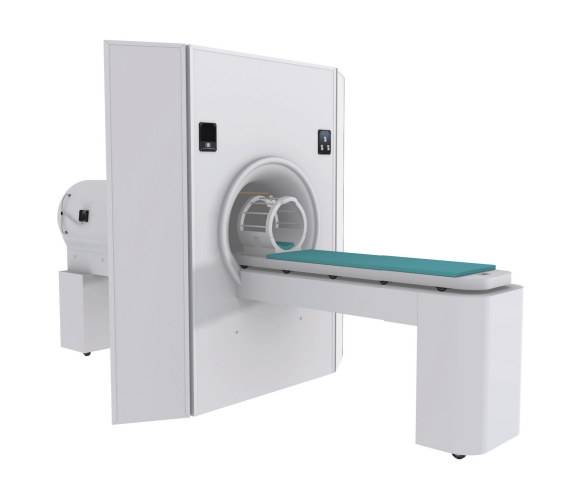
To acclimate patients to the MRI scanner experience, BIAC has recently purchased a new Vera MRI Simulator from Psychology Software Tools to replace the old mock scanner used in previous years. This new simulator includes an automated table, lights and fan, and provides an improved platform for potential subjects to experience what it will be like in the actual MRI machine. While the participant is in the mock scanner, you can display tasks to the subject, as well as play MRI sounds for various sequences. This system is a great resource to introduce participants to the identical experimental procedures that they will experience in the actual scanner, and to train children to participate in functional neuroimaging studies. This is done to ensure participant comfort and data quality.
 Below you will find some tips to help utilize and operate the mock scanner.
- The weight limit for the bed is 275 lbs.
- The power switch is located at the back of the simulator. After being turned on, it takes 2-3 minutes before the motor will engage and is ready to move the bed. Be patient. If left on for a long period of time (like overnight), it will also disengage, and you’ll need to turn it off and then back on.
- The control panel has buttons to operate the bed, the lights, and the fan, and are clearly marked.
- Pushing the button once will move the bed into the bore, it will stop when it reaches the end. A second press will stop the bed, the next press will move the bed out. It seems to always follow this sequence of move-stop-move-stop with each push, so if you send the bed all the way in, you may have to push the button a couple times to get it to come out (since the automatic stop doesn’t replace the push button stop in the sequence).
- On each side of the bed is an automatic safety stop. Pushing this disengages the motor, and you can pull the bed out (you can pull It out with the motor engaged, it’s just much harder).
Quality Assurance (QA)
BIAC routinely collects quality assurance data using AGAR phantoms on both 3T scanners. The goal is to collect this data on each scanner daily. At a minimum, it should be collected a few times per week. This data allows us to evaluate the scanners SNR and SFNR values, which in turn allow us to closely monitor scanner performance and quickly identify and diagnose problems. QA analysis tools also provide figures of merit for the mean signal, standard deviations, and signal stability in the X, Y, and Z planes. The QA tools BIAC has implemented with the support of the fBIRN are described (and can be downloaded) here: https://xwiki.nbirn.org:8443/bin/view/Function-BIRN/AutomatedQA
A history of regular QA can be seen here : BIAC-QA
In addition to BIAC's QA procedures, GE also runs a quality control program every month called ibis. This program monitors, and adjusts if necessary X, Y, and Z gradient shimming (LVshim). The GE system performance tests checks and logs stability and SNR data on the scanner, which can be recalled from the scanner's service browser. These procedures also test for coherent noise and eddy current problems.
Finally, for every scanning session, BIAC collects data on the SNR, SFNR, standard deviation, drift, mean signal, first time point of data (t1), and provides plots of signal stability in the X, Y , and Z planes (plot). These data are available to investigators immediately after the data has been collected and reconstructed. These can be found by logging into the BIAC Exam Tracker here: http://shadow.biac.duke.edu/biacweb/dashboard/ or here: http://www.biac.duke.edu/services/
Retired Equipment
GE EXCITE HD 3T Scanner (BIAC3 - Retired 9/6/2012)
The 3.0 T GE EXCITE HD scanner is a short bore magnet that uses an actively shielded magnet design to restrict fringe magnetic fields. This scanner is equipped with high-power 40 mT/m gradients at 150 T/m/s slew rate that enables single-shot echo-planar and spiral imaging at 64 x 64 and 128 x 128 matrices. The scanner uses a twin-gradient system, providing a 2.2 G/cm gradient for high-linearity imaging in body or large field of view (FOV) scans and a 4 G/cm gradient for a high-slew rate in fast imaging. The scanner also contains a parallel imaging infrastructure with an 8-channel head coil, with the option to upgrade to 16 channels.
The current software platform is 12xM4 (2/10/2010).
Whisperrooms
The BIAC has installed two Whisperroom sound-attenuating chambers (http://www.whisperroom.com/). These rooms are equipped with networked computers and 8 channel button boxes, identical to those used at the scanner. They are ideal for testing experimental paradigms and conducting behavioral studies. As they are located in close proximity to our MRI scanners, they are also often used for pre-scan training and post-scan testing.